Development of a Multispectral Collagen Detection Dermatology Device for Delineation of Non-Melanoma Skin Cancers
Advisor: Dr. Leonid Shmuylovich
Introduction & Motivation
Non-melanoma skin cancers (NMSCs), primarily basal cell carcinoma (BCC) and squamous cell carcinoma (SCC), are the most common cancers in the US. The gold standard for surgical clearance, Mohs micrographic surgery, relies on iterative, time-consuming histological assessments. In standard practice, dermatologists use their eyes or cross-polarized dermatoscopes to visually identify tumor margins prior to surgery. However, this subjective visual approach often fails to accurately determine subclinical tumor margins, leading to multiple surgical stages and prolonged procedures.
As NMSC tumors proliferate, they remodel the surrounding extracellular matrix, leading to a localized disruption of the dermal collagen network. Visualizing this collagen disruption can serve as an objective surrogate for tumor extent.
Multispectral Imaging & System Design
The overarching goal of this project is to create a practical, noninvasive imaging tool capable of guiding surgical excisions at the point of care. Prior multispectral systems required bulky external filters and motorized filter wheels, rendering them expensive and susceptible to motion artifacts.
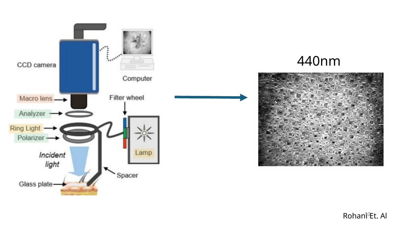
We designed a dual-wavelength snapshot system by repurposing a commercial cross-polarized dermatoscope. Our compact module utilizes a dichroic beamsplitter to divide the incoming light into two channels, captured simultaneously by two fixed monochrome cameras. A 450 nm blue band strongly detects collagen-dependent backscattering, while a 650 nm red band provides a reference sensitive to endogenous chromophores (like hemoglobin and melanin) but insensitive to collagen scattering. This design eliminates mechanical moving parts, minimizes inter-frame motion artifacts, and uses a Raspberry Pi for low-cost, portable control.
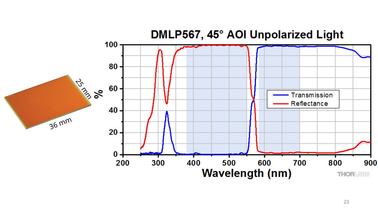
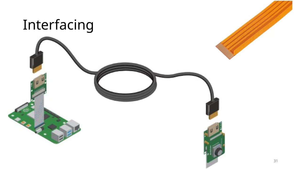
Image Analysis & Results
The raw 450 nm and 650 nm images are computationally registered to correct for minor spatial offsets introduced by the dichroic beamsplitter. By utilizing a spectral encoding formula, we generate a "collagen integrity map." This map isolates the loss of collagen scattering associated with NMSCs from other background variations.
The resulting scalar map is converted into an intuitive pseudo-color image for clinical interpretation, where regions of high collagen degradation appear in dark colors while intact collagen networks appear in warmer colors. By enhancing a tool dermatologists already trust, this low-cost, modular system has the potential to democratize advanced optical margin assessment, improving tumor clearance rates on the first surgical stage.

Device Design
Below are additional images and videos detailing the physical and optical architecture of the clinic-ready device prototype: